A general role for TANGO1, encoded by MIA3, in secretory pathway organization and function

Our latest work has now been published in Journal of Cell Science:

A general role for TANGO1, encoded by MIA3, in secretory pathway organization and function 

What’s more for the first time in ages, we have a cover. OK, the online cover. Still counts though. I won’t restate the paper here but instead will reflect on my favourite parts and why we don’t think our data support models where TANGO1 is selective for procollagen. Please do read the full paper for the background, detail, and greater context.

I have worked on the system governing the export of proteins from the endoplasmic reticulum in mammalian cells since 1999 and with the team in Bristol since 2001. While I think our lab has made some significant contributions, we have of course always followed the field with great interest. A part we have always been reluctant to tackle from has been TANGO1, encoded by Mia3. Initially reported as a co-factor for procollagen secretion in 2009, substantial data since then has elaborated this role to include some other large cargoes, more nuanced mechanisms involving engagement of Hsp47 (a key chaperone) rather than procollagen itself, and a broader role at the point of ER exit through recruitment of a tethering complex located at the ER-Golgi Intermediate compartment (ERGIC) the first post-ER compartment. You can read up on work to date regarding the ER-Golgi interface and work to date in our review article in Trends in Cell Biology.

With the advent of CRISPR-Cas9 gene editing and Janine McCaughey’s inexhaustible thirst for new data, we embarked on a project to edit the genome of human cells to try and eliminate expression from the Mia3 gene that includes TANGO1. The paper goes into much more depth here of course but the key issue is that there are two major (and likely several minor) isoforms of TANGO1, the shorter of which has only a small region within the lumen of the ER. This has previously been shown (by RNAi and other work) to be sufficient to support secretion of procollagen.

The most effective “knockout” that we have almost (but not quite) eliminates expression fo all isoforms. This showed a dramatic phenotype (which made the cover) of a multitude of vesicular structures at the interface of the ER and Golgi.

Cover of the issue of Journal of Cell Science in which the article is published
Transmission electron microscopy image of a cell with disrupted ER-to-Golgi trafficking following CRISPR-Cas9-mediated editing of MIA3/TANGO1.

I love this image as it is so thought-provoking and generates ideas every time I look at it. My own speculation here (I use that word advisedly based on feedback!) is that TANGO1 constrains the formation of COPII-coated vesicles to generate a functional ERGIC. In its absence, COPII vesicle formation is more efficient (maybe like in other organisms that don’t express TANGO1 or really have a definable ERGIC????) and that leads to this phenotype. Trafficking works perfectly well in organisms that don’t have TANGO1 but they maybe don’t have the same secretory load as vertebrates. So, I see TANGO1 as an “optimiser” of the system, not as a de facto procollagen/large cargo packaging factor.
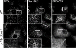
Image showing how loss of TANGO1 expression affects the ERGIC
Immunofluorescence image from the paper showing collapse of the ERGIC on loss of TANGO1 expression

In my mind I see this most closely aligned to the data in the paper that shows a “collapse” of the ERGIC with markers ERGIC53 and SURF4 becoming exclusively localized to the ER. my interpretation – we have lost the ability to either build or maintain a functional ERGIC. Maybe these vesicles represent a fragmented Golgi? Maybe (and we’re trying some immuno-EM of course) but this phenotype doesn’t look like a COPI block or other Golgi-based perturbations that I have seen.

Why does this matter? Many organisms get along fine without a definable ERGIC (but then many of these same organisms get along fine with a very differently organization of the Golgi). The previous data on the role of TANGO1 in recruiting the ERGIC becomes key here. What if in fact its role is to support biogenesis of the ERGIC? What if the role of the ERGIC is to support efficient cargo secretion? Any loss of integrity here would lead to defects in secretion which we see in multiple assays in the paper, including proteomics.

The impact on secretion here is significant. Surely if we block ER export then this is what you would expect to see? Well, ER export isn’t blocked. Indeed, our proteomics data show enhanced secretion of some cargoes. For the majority of “canonical” cargoes secretion is slowed. This is also true for procollagen which is seen to accumulate inside cells (the “L” lanes on the blot below).

Immunoblots showing defective procollagen secretion

Loss of TANGO1 causes procollagen to accumulate inside cells. The severity of the defect correlates with the severity of disruption to the Mia3 locus. So where does this leave us in terms of our interpretation that TANGO1 is NOT procollagen- or “large cargo”-selective?

My favoured model here is that procollagen is one of the most significantly impacted cargoes – defects in its secretion are seen on perturbation of almost any key component of the early secretory pathway from COPII subunits (e.g. Sec13 and Sec23), components of ER-Golgi tethers e.g. Sedlin and NBAS, and Golgi proteins such as GMAP210, giantin, and GORAB.

In our data we show that many other cargoes are affected, small soluble proteins, larger matrix proteins, and transmembrane proteins. We’ve used the RUSH system to retain and then control release of cargo (including procollagen) from the ER – this leads to the critique that we are overexpressing and that we are forcing a large synchronous cargo load. Fair enough but we have back up these data with other methods – the blot here is on endogenous procollagen, proteomics of the soluble “secretome” from these cells as well as of the cell-derived matrix show major changes in secretion.

Another key factor we find with regard to reduced secretion of collagen in general is that the expression of many collagens is downregulated. The RNAseq data are a key component of this work and one that we have yet to explore fully. Type XI collagen is strongly affected here and is critical for the formation of a fibrillar matrix. This could impact key defects seen with in vivo mouse models and in humans (here and here) with disrupted Mia3 expression. We do not underestimate the likely impact of changes in gene expression to the phenotypes we see.

Importantly we can also restore key phenotypes with the short form of TANGO1 which suggests that engagement with Hsp47/procollagen might not be required for at least some of this.

I reflect back to our reviews on this particularly the one in Trends in Cell Biology where we discuss the importance of the ERGIC as a “buffer” to maintain compartment identity of the ER and the Golgi. I think there is also now more of a consensus that a more amorphous membrane network traffics cargo through the early secretory pathway and our contention is that TANGO1 is a key component that sets up this membrane network. Much of this also echoes back to work, notably from Hans-Peter Hauri (now retired) on the identity of the ERGIC. Our data indicate that it is not a stable compartment but requires TANGO1 for its existence – whether this is really in terms of ERGIC biogenesis or maintenance remains to be defined.

This of course is not the whole story by any means. We are now developing this work thanks to further UKRI-BBSRC funding and will be able to explore phenotypes in vivo thanks to a new collaboration with Brian Link at the Medical College of Wisconsin who has made several zebrafish models of relevance here. We hope that the proteomics and genomics data in this paper will inform much of this work. We also of course make that data freely available to all, such that anyone can build on this study. Plasmids are also in Addgene for those interested.

I hope you will read the full paper. As usual – a great experience publishing with Journal of Cell Science (COI – I am an Editor but it really is a great experience!).

New data: Giantin is required for 1 intracellular N-terminal processing of type I procollagen

Our new preprint is now available on bioRxiv. We are really excited to share this new work from Nicola Stevenson which comes from our great collaboration with Chrissy Hammond (@ChrissyLHam) and Dylan Bergen (@SciBergen)  https://www.biorxiv.org/content/10.1101/2020.05.25.115279v1

Some background:

Giantin is a golgin, it is ubiquitously expressed and conserved throughout vertebrates at least. Golgins define the specificity of incoming vesicle trafficking to the Golgi (beautiful work from Mie Wong and Sean Munro (Wong and Munro, 2014 https://science.sciencemag.org/content/346/6209/1256898/tab-article-info).

However, in their work, they have not been able to assign a role for giantin as a vesicle tether. Our own work has shown that loss of giantin results in minor defects in Golgi organization but is required for normal gene expression of some glycosyltransferases, notably GALNT3. doi: 10.1242/jcs.212308

It is also required for normal cilia formation in cells (Asante et a., 2013; Stevenson et al., 2017, Bergen et al., 2017)
(https://jcs.biologists.org/content/126/22/5189 and https://jcs.biologists.org/content/131/9/jcs212258) and zebrafish (https://bio.biologists.org/content/6/8/1180).

Various model organisms with mutations in or deletions of giantin, including our zebrafish models, show consistent defects in extracellular matrix formation. We sought to define this in more detail using our own zebrafish mutants and giantin KO cell lines.

The new data:

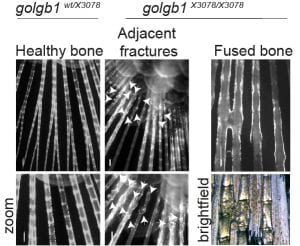
We find that giantin mutant zebrafish are prone to spontaneous fractures and bone mineralization defects. There is also a small but reproducible reduction in expression of type I procollagen in these tissues.

To look at this in more detail we switched to our giantin KO cells where we had also identified a consistent decrease in COL1A1 gene expression. Despite that, to our surprise we found a clear INCREASE in type I procollagen protein in these cells.

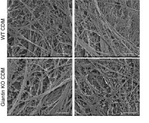
This was accompanied by increased secretion of type I procollagen and small but clear defects in the organization of extracellular matrix derived from these cells.

Using live cell imaging, we were surprised to find that these cells showed no apparent differences in trafficking of pro-α1(I) compared to controls.

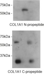
Now we come to the most exciting part for me – what we did find was a clear defect in N-terminal processing of the N-terminal propeptide of pro-α1(I). Specifically, that this part of the precursor is NOT effectively cleaved in giantin KO cells.

N-pro is retained until pro-α1(I) reaches the extracellular space. Most significantly, the N-propeptide is readily detected in cell lysates of WT cells but not in giantin KO cells. Our GFP-tagged collagen that we have used to define the intracellular trafficking route reports this really effectively using a variety of tags.

No defects were seen in processing of the C-terminal propeptide.

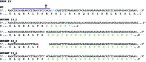
This finding was robustly reproduced in Nikki’s experiments and indeed, because of possible issues around clonal selection of our KO cells and the fact we only had one line, Nikki then generated further giantin KO lines.

Really intriguingly, these all show procollagen processing defects but each in a slightly different way. This supports our idea that this is not a direct mechanistic link between giantin and procollagen processing but rather reflects a fundamental defect at the Golgi.

We have spent years chasing the N-pro enzymes, especially the ADAMTS proteins but we haven’t found antibodies that detect them robustly nor have we been able to generate any tagged forms of ADAMTS2/3/14 that leave the ER.

There is also a lot of other data in here and mentions of what hasn’t worked or defined clear outcomes. Gaps and questions aplenty!

Can we define a direct link between giantin and the processing machinery? No. That said, we have to question whether our current understanding of ADAMTS biology is accurate (what if they are in fact primarily localized to the ER?).

We also can’t prove that the bone defects including susceptibility to fracture is directly linked to the N-pro defect. This is primarily because we can’t detect pro-α1(I) by immunoblotting in fish.

So what do we know from this?

  • Giantin is required for normal bone integrity in zebrafish
  • Giantin is also required for normal N-terminal propeptide cleavage of pro-α1(I)

Relationship to other work:

There is precedent for detection of intracellular procollagen processing in both chick and mouse tendons (Canty‐Laird et al., 2012; Canty et al., 2004; Humphries et al., 2008). Here, Kadler and colleagues found processed forms of type I procollagen in a detergent soluble fraction from tendon explants, consistent with an intracellular pool. This is however, also consistent with a detergent soluble extracellular pool.

In other experiments, his lab showed the presence of the N-propeptide in the presence of brefeldin A (which causes merging of the Golgi with the ER) (Canty-Laird et al., 2012). We consider that this shows clearly that the N-pro “can” be cleaved inside cells when you force the enzyme and substrate together using BFA.

We consider that our data shows that giantin is needed for this cleavage event to occur inside cells. This is the first defined “function” for giantin and clear support for the concept of intracellular processing of type I procollagen.

So why don’t we see a clearer phenotype in giantin mutant fish? Why doesn’t this reflect an EDS type VII phenotype (which arises from mutations in the N-pro enzyme)? The most likely explanation is that extracellular processing can occur but is inefficient.

If that is all true, then why would some pro-α1(I) be cleaved inside the cell if all can be cleaved outside? This raises the possibility of different pools of procollagen that are cleaved at different locations or at different times.

Could this relate to the ultimate role of that collagen? Maybe tissue formation versus repair? Could this relate to circadian control of matrix composition? We don’t know but we’re working on these and more ideas right now as part of our BBSRC-funded work with Karl and his colleagues in Manchester.

Is this work “perfect”? No. Is this complete? No. BUT we can say it is really robust, provides some key insight that will hopefully allow us and others to build on to better understand collagen matrix assembly and repair.


References:

Asante, D., L. Maccarthy‐Morrogh, A.K. Townley, M.A. Weiss, K. Katayama, K.J. Palmer, H. Suzuki, C.J.
620 Westlake, and D.J. Stephens. 2013. A role for the Golgi matrix protein giantin in ciliogenesis through
621 control of the localization of dynein‐2. J Cell Sci. 126:5189‐5197.

Bergen, D.J.M., Stevenson, N.L., Skinner, R.E.H., Stephens, D.J. and Hammond, C.L. (2017)

The Golgi matrix protein giantin is required for normal cilia function in zebrafish

Canty‐Laird, E.G., Y. Lu, and K.E. Kadler. 2012. Stepwise proteolytic activation of type I procollagen to
648 collagen within the secretory pathway of tendon fibroblasts in situ. Biochem J. 441:707‐717.

Canty, E.G., Y. Lu, R.S. Meadows, M.K. Shaw, D.F. Holmes, and K.E. Kadler. 2004. Coalignment of plasma
652 membrane channels and protrusions (fibripositors) specifies the parallelism of tendon. J Cell Biol.
653 165:553‐563.

Humphries, S.M., Y. Lu, E.G. Canty, and K.E. Kadler. 2008. Active negative control of collagen fibrillogenesis
694 in vivo. Intracellular cleavage of the type I procollagen propeptides in tendon fibroblasts without
695 intracellular fibrils. J Biol Chem. 283:12129‐12135.

Stevenson, N.L., D.J.M. Bergen, R.E.H. Skinner, E. Kague, E. Martin‐Silverstone, K.A. Robson Brown, C.L.
779 Hammond, and D.J. Stephens. 2017. Giantin‐knockout models reveal a feedback loop between Golgi
780 function and glycosyltransferase expression. J Cell Sci. 130:4132‐4143.

Wong, M., and S. Munro. 2014. Membrane trafficking. The specificity of vesicle traffic to the Golgi is
793 encoded in the golgin coiled‐coil proteins. Science. 346:1256898.

Skip to toolbar